

Cartilage Ulcers
Introduction/ What is?
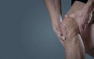
Joints in our bodies comprise ends of bones articulating with each other. The ends of the bones forming the joints are covered with an extremely smooth cartilage further lubricated with viscous joint fluid, making for friction-free movements. Holes in the smooth cartilage cap can occur at any joint resulting. These holes are known as cartilage ulcers.

What are the symptoms?
The common symptoms of cartilage ulcers are:
- Dull ache of the knee after activities
- Swelling of the knee
- Pain with certain movements of the knee especially when bearing weight (eg squatting or running)
- Popping, clicking or grating sensation in the knee with movements
What caused it?
Cartilage ulcers usually occur due to “overuse” during sports and symptoms develop gradually. Sometimes, however, they can occur if the particular joint suffers a sudden high energy injury like a violent twisting or yanking injury during sports.
Can it heal?
Yes, some cartilage ulcers can heal. How, not all do. It is not possible to predict which ones will heal. Certainly if pain and symptoms have been around for a long time (months), the likelihood of it healing are low.
What tests can be done to diagnose this?
An MRI scan is by far the best way to detect presence of a cartilage ulcer.
What treatment options are there?
Small, superficial ulcers can be left alone and treated with anti-inflammatory medications and physiotherapy. Small, full thickness cartilage ulcers which are not overly symptomatic can also be treated similarly.
Larger, full thickness ulcers often require more attention in the form of various injections or surgery. Injections are generally not effective in bringing about healing alone, but may be administered as an adjunct to surgery.
There are many cartilage repair surgeries. Most are performed via keyhole surgery. It is best to speak to your orthopaedic doctor to explore the most suitable option for yourself.

 ">
">
 Dr Bryan Tan